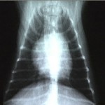
 Diagnostic imaging in pets has improved tremendously just as it has for people. There are now the same tests available to our dogs and cats as for us; including radiography (x-rays), ultrasound as well as MRI and CT scans. Each of these tests has its own advantages and disadvantages and provides vastly different information.
Here at the Dog & Cat Clinic of Niagara we offer both digital radiographs and ultrasound. If needed, we also can refer you and your pet to referral facilities that offer MRI and CT scans.
A radiograph is a black and white two-dimensional picture of the inside of the body. This picture is generated by passing radiation through the body area to be investigated. The traditional way of recording the picture is on a specific x-ray film that captures the radiation much like the way photographic film captures light. Less dense structures such as air in the lungs allow most of the x-rays to pass through to the film, turning the area black. Bone stops the radiation from reaching the film and thus appears white. All of the other structures are varied shades of gray depending on their density.
What is “Digital” Radiography? The principals are the same as traditional radiographs but x-ray film is not used. Instead, the images are captured on a digital recording device. This form of x-ray uses much less radiation thus making it safer for your pet. The images are also processed faster allowing a more efficient diagnostic process. These digital images are also easy to store directly within your dog or cat’s computerized medical records. These records and images can easily be sent over the internet to other veterinarians including our emergency after-hours service and specialists allowing a cohesive approach to the care of your pet.
